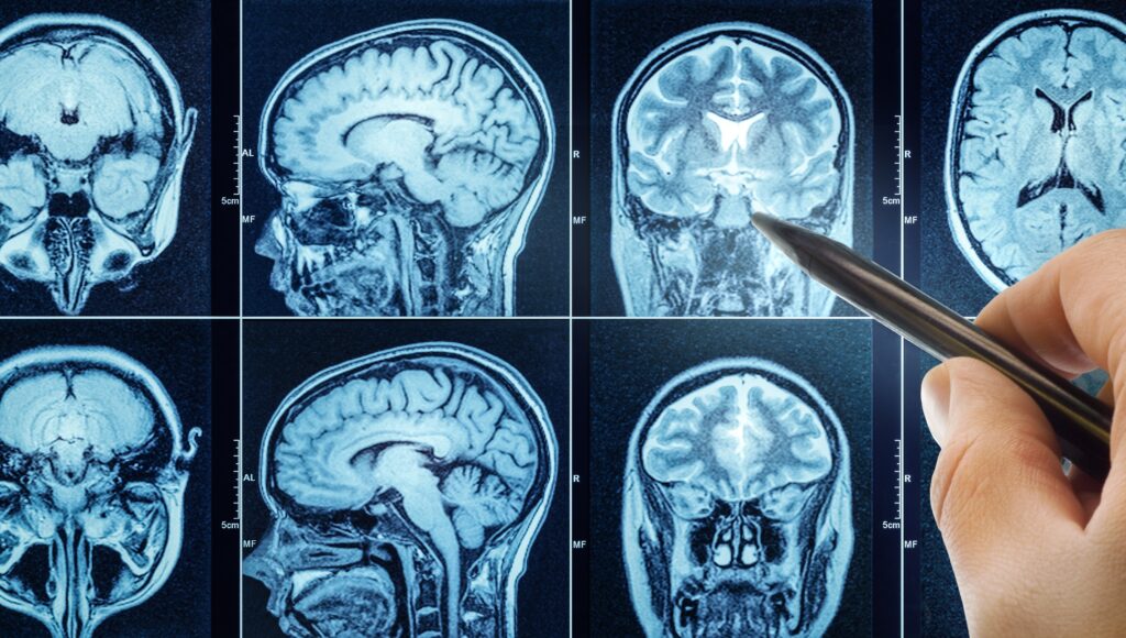
Researchers in the UK have made a significant breakthrough by using live human brain tissue to recreate the early stages of Alzheimer’s disease. The study, conducted at the Royal Infirmary of Edinburgh, involved exposing healthy brain cells from living patients to a toxic form of amyloid beta, a protein closely linked to Alzheimer’s. By studying this process in real-time, scientists were able to observe how Alzheimer’s damages the brain, which could accelerate the development of more effective treatments for the disease.
Brain Tissue Collected During Surgery and Kept Alive in the Lab
The groundbreaking research involved brain tissue collected from patients undergoing tumor removal surgeries. These surgeries took place at the Royal Infirmary of Edinburgh, where small, healthy fragments of brain tissue were carefully removed and given to researchers during the procedure. Normally, such tissue would be discarded, but in this case, it was kept alive in the lab for further study.
The collected tissue was immediately placed in oxygen-rich artificial spinal fluid and transported to a nearby laboratory. There, researchers sliced the tissue into thin sections and maintained it in nutrient-rich fluid at body temperature. The team began experiments immediately, with the tissue remaining alive for up to two weeks, thanks to patient consent. This allowed the team to study the effects of amyloid beta in real-time.
Toxic Amyloid Beta Causes Irreversible Damage
To recreate the conditions of Alzheimer’s disease, researchers applied toxic amyloid beta to the tissue. Unlike the normal, non-toxic version of the protein, the toxic form caused significant damage that the brain cells could not repair. Even slight changes in the amyloid beta levels led to disrupted cell functions, showing that the brain requires precise levels of this protein for normal operation. This finding is crucial because it highlights the role of amyloid beta in the early stages of Alzheimer’s and provides insight into how the disease progresses.
New Insights into Vulnerable Brain Regions and Disease Progression
The study also revealed new clues about which brain regions are most vulnerable to Alzheimer’s. Dr. Claire Durrant and her team discovered that brain tissue from the temporal lobe—an area often affected early in Alzheimer’s disease—released higher amounts of tau protein. Tau is another protein that plays a central role in the development of Alzheimer’s. The higher levels of tau in the temporal lobe may explain why this region is impacted early on in the disease. Researchers suggest that the accumulation of tau protein could help the toxic amyloid beta spread more quickly from cell to cell, accelerating the progression of the disease.
Support for the Research
This innovative research was supported by Race Against Dementia and received a £1 million donation from the James Dyson Foundation. Dyson, who is known for his work in engineering and innovation, praised the use of live human brain tissue for this study, noting that it is a far more accurate model than traditional animal studies. The research aims to provide a clearer understanding of how Alzheimer’s progresses in human brains and offers a platform to test potential treatments directly on human brain cells.
Professor Tara Spires-Jones, a leading researcher in the field, emphasized the importance of the study in helping scientists better understand the disease. “This research will allow us to observe Alzheimer’s progression in real-time and test new therapies that could ultimately lead to more effective treatments for patients,” she said.
Implications for Alzheimer’s Treatment
This research is expected to have significant implications for the future of Alzheimer’s treatment. By observing the effects of amyloid beta and tau on human brain tissue, scientists can gain valuable insights into the mechanisms behind the disease. This method could also pave the way for testing new drugs and therapies directly on human brain cells, potentially speeding up the process of finding effective treatments.
The use of live human brain tissue represents a significant step forward in Alzheimer’s research, as it allows for a more accurate understanding of the disease’s early stages and progression. With continued support and further studies, this breakthrough could contribute to a much-needed advancement in the search for a cure for Alzheimer’s.
The groundbreaking study using live human brain tissue offers a new and more accurate approach to studying Alzheimer’s disease. By recreating the early stages of the disease, researchers can now observe how amyloid beta and tau affect brain cells in real-time, providing new clues about disease progression and vulnerable brain regions. With continued research and the development of more effective testing methods, this breakthrough could ultimately lead to better treatments for Alzheimer’s disease and a deeper understanding of how to combat it.
